Matière et Systèmes Complexes
Présentation
Le laboratoire « Matière et Systèmes Complexes » (MSC) est une unité mixte de recherche du CNRS et de l’université (UMR 7057). Le laboratoire est installé depuis 2007 sur le nouveau campus de l’Université Paris Diderot, Paris Rive Gauche, dans le bâtiment Condorcet. Il est réparti sur plusieurs étages. La direction et le secrétariat se trouvent au 6e étage. Le directeur actuel en est Laurent Limat, secondé par la directrice adjointe Florence Gazeau.
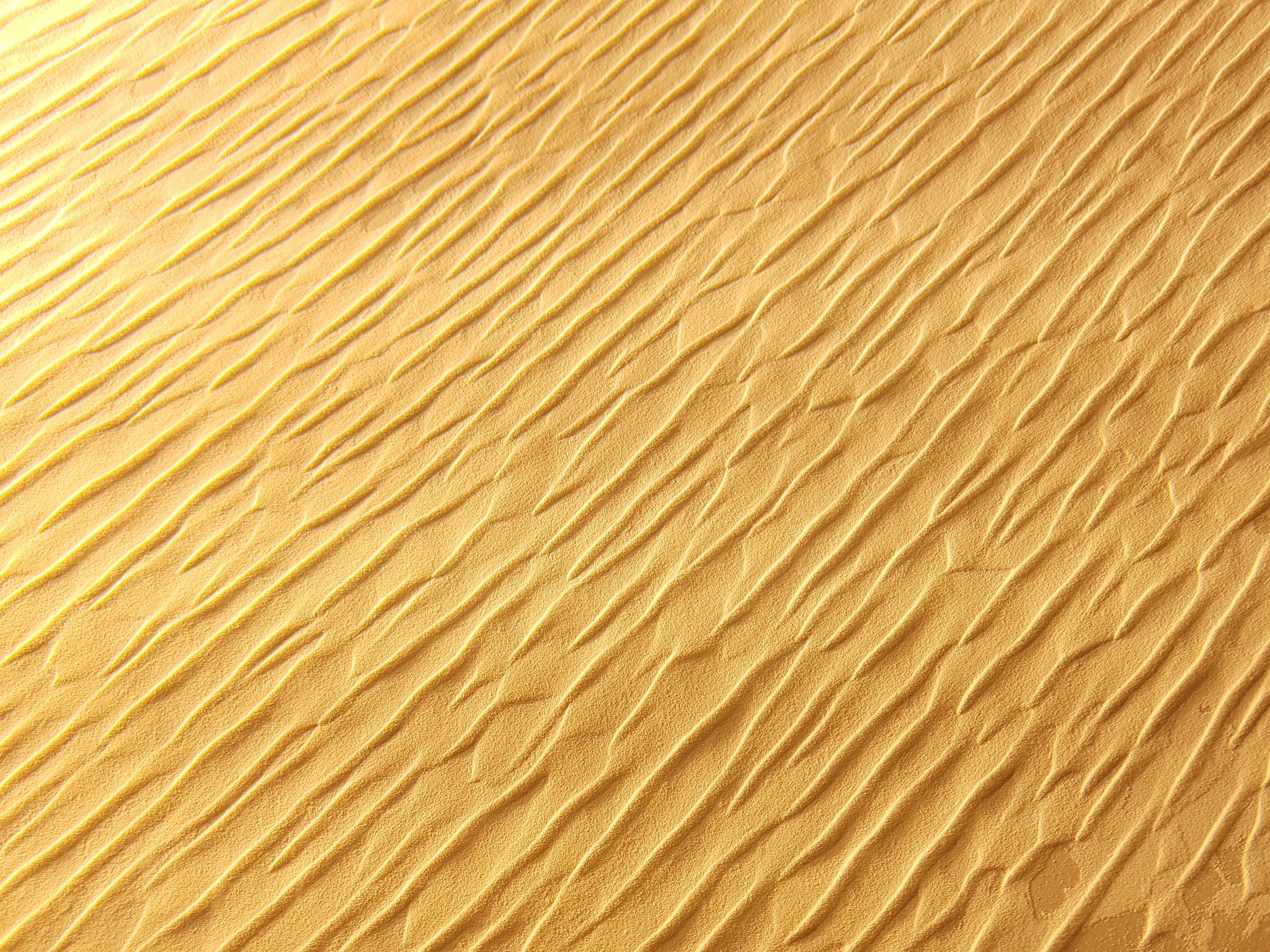
Le laboratoire MSC a pour sujet d’étude la matière et les systèmes complexes sous toutes leurs formes. Il peut s’agir de fluides montrant des phénomènes complexes non-linéaires (facettages de jets ou de tourbillons, structures et propriétés complexes de mousses, phénomènes de mouillage, propagation de vagues et de tsunamis) ou bien, par exemple, de systèmes proches de la géophysique et de l’environnement (systèmes granulaires tels que les dunes, phénomènes d’érosion, morphogenèse des plantes et même des villes, nage collective d’algues ou de bactéries…). Les études théoriques et expérimentales conduisent à des applications comme par exemple les éoliennes flexibles de haut rendement, l’optimisation de méthodes d’enduisage, le contôle de propriétés de surface ou la récupération de la biomasse (ingénierie verte)...
Le laboratoire étudie également le couplage entre la physique et la biologie des systèmes vivants, avec une approche multi-échelle. Les recherches effectuées vont d’échelles moléculaires ou supra-moléculaires (assemblages des protéines, chromatine, cytosquelette etc.) jusqu’à l’échelle de l’organisme entier (méduses, poulets, vers etc.) en passant par des études plus fondamentales sur des cellules uniques sur lesquelles sont exercées des forces quantifiées, permettant de comprendre les propriétés biophysiques de la matière vivante. Ces études aboutissent à de possibles applications en ingénierie tissulaire ou régénération des tissus avec des transferts dans le domaine médical.
Equipes de recherche
Le laboratoire est structuré en cinq équipes :
- Dynamique des systèmes hors d’équilibre (DSHE), orientée plutôt vers les comportements non-linéaires de fluides, éventuellement actifs ou avec surface libre, et les phénomènes d’auto-organisation en général (morphogenèse des granulaires, systèmes particulaires inspirés de la matière condensée, colloïdes et transition d’encombrement, etc).
- Dynamique et organisation de la matière molle (DOMM), orientée plutôt vers les matériaux mous visco-élastiques aux propriétés rhéologiques complexes (gels, polymères, mousses etc.), milieux caractérisés par une structure hétérogène, et dont l’organisation et les propriétés dépendent de l’échelle d’observation.
- Physique du vivant, orientée plutôt vers l’étude des processus physiques qui sous-tendent les fonctions biologiques, principalement à l’échelle cellulaire, entre la molécule et le tissu.
- Biofluidique, orientée plutôt vers l’étude des systèmes vivants du tissu à l’organisme, avec des applications à visées médicales.
- Une équipe de théoriciens dont les thématiques couvrent un spectre large de questions fondamentales allant de la physique statistique hors équilibre à la neuroscience, en passant par la matière molle et la matière active.
Cependant les activités de ces équipes se recoupent souvent dans des projets communs aux frontières entre les comportements physiques et/ou biologiques (exemple : comportement de mousses marines, mesures de forces dans des tissus reconstitués, etc.)
[hal-00112538] Zipper Dynamics of Surfactant Nanotube Y Junctions
Date: 9 Nov 2006 - 09:30
Desc: We investigate the formation of Y junctions in surfactant nanotubes connecting vesicles. Based on experimental observations of the surfactant flow on the nanotubes, we conclude that a Y junction propagates with a zipperlike mechanism. The surfactants from two nanotube branches undergo 1∶1 mixing at the junction, and spontaneously form the extension of the third nanotube branch. Taking into account the tension driven surfactant flow, we develop a model for the Y junction dynamics that is in quantitative agreement with the experimental data.
[hal-02463702] Mechanical Control of Morphogenesis by Fat/Dachsous/Four-Jointed Planar Cell Polarity Pathway
Date: 1 Feb 2020 - 15:48
Desc: [...]
[hal-00005362] Microfabricated arrays of elastomeric posts to study cellular mechanics
Date: 14 Jun 2005 - 16:49
Desc: We present an approach to fabricate an array of elastomer posts in order to dynamically measure the traction forces exerted by living cells on a surface with a micrometer lateral resolution. Arrays of closely spaced vertical microposts are made in silicone elastomer [poly(dimethylsiloxane) (PDMS)] by molding a Silicon substrate that has been machined by deep Si etching after standard photolithography. The surface of the micropillars was modified to allow cell culture. Deflections of the calibrated posts were dynamically followed by direct obervation with an optical microscope. By using this set-up, we could dynamically draw up a cartography of the local traction forces exerted by the cells.
[hal-00175727] Collective migration of an epithelial monolayer in response to a model wound
Date: 1 Oct 2007 - 10:24
Desc: Using an original microfabrication-based technique, we experimentally study situations in which a virgin surface is presented to a confluent epithelium with no damage made to the cells. Although inspired by wound-healing experiments, the situation is markedly different from classical scratch wounding because it focuses on the influence of the free surface and uncouples it from the other possible contributions such as cell damage and/or permeabilization. Dealing with Madin–Darby canine kidney cells on various surfaces, we found that a sudden release of the available surface is sufficient to trigger collective motility. This migration is independent of the proliferation of the cells that mainly takes place on the fraction of the surface initially covered. We find that this motility is characterized by a duality between collective and individual behaviors. On the one hand, the velocity fields within the monolayer are very long range and involve many cells in a coordinated way. On the other hand, we have identified very active “leader cells” that precede a small cohort and destabilize the border by a fingering instability. The sides of the fingers reveal a pluricellular actin “belt” that may be at the origin of a mechanical signaling between the leader and the followers. Experiments performed with autocrine cells constitutively expressing hepatocyte growth factor (HGF) or in the presence of exogenous HGF show a higher average velocity of the border and no leader.
[hal-03761686] Strength Dependence of Cadherin-Mediated Adhesions
Date: 26 Aug 2022 - 13:17
Desc: Traction forces between adhesive cells play an important role in a number of collective cell processes. Intercellular contacts, in particular cadherin-based intercellular junctions, are the major means of transmitting force within tissues. We investigated the effect of cellular tension on the formation of cadherin-cadherin contacts by spreading cells on substrates with tunable stiffness coated with N-cadherin homophilic ligands. On the most rigid substrates, cells appear well-spread and present cadherin adhesions and cytoskeletal organization similar to those classically observed on cadherin-coated glass substrates. However, when cells are cultured on softer substrates, a change in morphology is observed: the cells are less spread, with a more disorganized actin network. A quantitative analysis of the cells adhering on the cadherin-coated surfaces shows that forces are correlated with the formation of cadherin adhesions. The stiffer the substrates, the larger are the average traction forces and the more developed are the cadherin adhesions. When cells are treated with blebbistatin to inhibit myosin II, the forces decrease and the cadherin adhesions disappear. Together, these findings are consistent with a mechanosensitive regulation of cadherin-mediated intercellular junctions through the cellular contractile machinery.
Autres contacts
Université Paris Diderot - Paris 7
U.F.R. Physique
Bâtiment Condorcet
10, rue Alice Domon et Léonie Duquet
75205 PARIS CEDEX 13