The Laboratory of Theoretical Biochemistry (LBT) is one of five laboratories within Institut de Biologie Physico-Chimique (IBPC) in Paris.
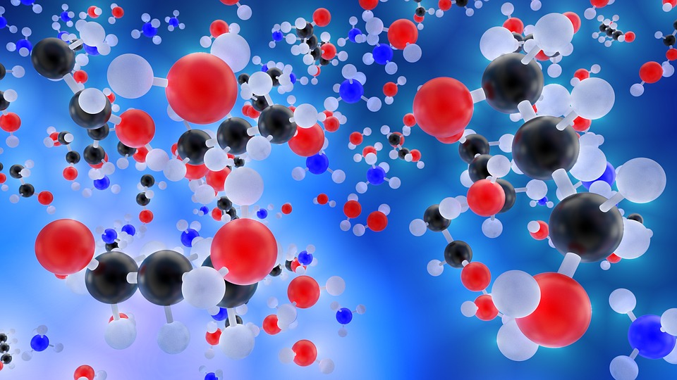
LBT belongs to the French national research agency CNRS through its Institute of Chemistry, and is associated with Paris Diderot University. The laboratory was created at IBPC in 1958 as Laboratoire de Biochimie Théorique. Our field is theoretical and computational biochemistry, at the interface between biology, chemistry, physics, and computing.
Our strategic objectives are twofold: invent simulation algorithms to reproduce and predict physical properties of biomolecules either in vitro or in the cell, and understand the molecular or conformational factors responsible for the biological functions of living systems, and diseases. The equilibrium between these two aspects is the key point of the laboratory policy.
LBT is organized as a team of independent researchers with complementary interests and domains of expertise, both in method development and in biophysical, biochemical, and biomedical applications. Advances in each of these domains emerge from the association of different sets of researchers around individual projects.
Les axes de recherches du LBT se concentrent sur les développements méthodologiques et algorithmiques pour l’étude de la structure, la dynamique, la mécanique et les interactions des macromolécules biologiques.
Les objectifs sont donc d'utiliser les ordinateurs pour ouvrir des fenêtres vers le monde moléculaire, en aidant à comprendre les facteurs qui sous-tendent des faits expérimentaux, et en prédisant les propriétés et le comportement des molécules biologiques.
Directeur : Marc Baaden
Date: 10 nov 2020 - 18:12
Desc: Phosphorylation of Na V 1.5 channels regulates cardiac excitability, yet the phosphorylation sites regulating channel function and the underlying mechanisms remain largely unknown. Using a systematic quantitative phosphoproteomic approach, we analyzed Na V 1.5 channel complexes purified from non-failing and failing mouse left ventricles, and we identified 42 phosphorylation sites on Na V 1.5. Most sites are clustered, and three of these clusters are highly phosphorylated. Analyses of phosphosilent and phosphomimetic Na V 1.5 mutants revealed the roles of three phosphosites in regulating Na V 1.5 channel expression and gating. The phosphorylated serines-664 and -667 regulate the voltage-dependence of channel activation in a cumulative manner, whereas phosphorylation of the nearby serine-671, which is increased in failing hearts, decreases cell surface Na V 1.5 expression and peak Na + current. No additional roles could be assigned to the other clusters of phosphosites. Taken together, the results demonstrate that ventricular Na V 1.5 is highly phosphorylated, and that the phosphorylation-dependent regulation of Na V 1.5-encoded channels is highly complex, site-specific and dynamic.
Date: 29 Mar 2017 - 17:06
Desc: The molecular mechanism underlying channel opening in response to agonist binding remains a challenging issue in neuroscience. In this regard, many efforts have been recently undertaken in ATP-gated P2X receptors. Among those efforts, we have provided evidence in the P2X2 receptor that tightening of ATP sites upon agonist binding induces opening of the ion channel. Here we extend our analysis to show that the sulfhydryl-reactive ATP analog 8-thiocyano-ATP (NCS-ATP), a potent P2X2 agonist, when covalently labeled in the ATP-binding site at position Leu186 likely favors the tightening mechanism, but not the channel opening mechanism. Our data predict the existence of intermediate or preactivation state(s) trapped by NCS-ATP, in which tightening of the binding site is favored while the channel is still closed. We propose that this (these) intermediate ATP-bound state(s) prime(s) channel gating in the P2X2 receptor.
Date: 7 oct 2019 - 11:48
Desc: P2X receptors (P2XRs) are ligand-gated ion channels activated by extracellular ATP. Although the crystal structure of the zebrafish P2X4R has been solved, the exact mode of ATP binding and the conformational changes governing channel opening and desensitization remain unknown. Here, we used voltage clamp fluorometry to investigate movements in the cysteine-rich head domain of the rat P2X1R (A118-I125) that projects over the proposed ATP binding site. On substitution with cysteine residues, six of these residues (N120-I125) were specifically labeled by tetramethyl-rhodamine-maleimide and showed significant changes in the emission of the fluorescence probe on application of the agonists ATP and benzoyl-benzoyl-ATP. Mutants N120C and G123C showed fast fluorescence decreases with similar kinetics as the current increases. In contrast, mutants P121C and I125C showed slow fluorescence increases that seemed to correlate with the current decline during desensitization. Mutant E122C showed a slow fluorescence increase and fast decrease with ATP and benzoyl-benzoyl-ATP, respectively. Application of the competitive antagonist 2',3'-O-(2,4,6-trinitrophenyl)-ATP (TNP-ATP) resulted in large fluorescence changes with the N120C, E122C, and G123C mutants and minor or no changes with the other mutants. Likewise, TNP-ATP-induced changes in control mutants distant from the proposed ATP binding site were comparably small or absent. Combined with molecular modeling studies, our data confirm the proposed ATP binding site and provide evidence that ATP orients in its binding site with the ribose moiety facing the solution. We also conclude that P2XR activation and desensitization involve movements of the cysteine-rich head domain.
Date: 29 Mar 2017 - 17:04
Desc: We review foundations of biomolecular simulations that enable the study of membrane protein models with a particular focus on structure–function relationships and opportunities for drug design. A range of broadly used methods is presented comprising homology modeling, normal mode analysis, molecular dynamics simulations, and free energy calculations. These methods are illustrated with examples on several membrane protein systems, in particular ligand-gated ion channels such as the P2X receptors, the N-methyl-D-aspartate (NMDA) receptors, and the Cys-loop family of pentameric ion channels.
Date: 29 Mar 2017 - 17:04
Desc: no abstract
Institut de Biologie Physico-Chimique (IBPC)
13, rue Pierre et Marie Curie
75252 PARIS CEDEX 05